Ultrasound imaging improves safety in breast fat transfer by allowing the surgeon to see beneath the skin in real time, so each small deposit of fat is placed in the intended tissue layer. This visual confirmation supports more controlled and precise enhancement of the breasts with your own tissue.
In breast fat grafting, fat taken from another area of your body is placed into the breast in many small amounts. The breast sits over the chest wall, with several distinct tissue layers in a small space. Where each deposit goes matters both for safety and for how well the graft survives.
Traditionally, surgeons have judged depth by anatomy, experience and the feel of the instrument. Those skills remain essential. Ultrasound adds a second layer of confirmation: the surgeon can see the layer, not only feel for it.
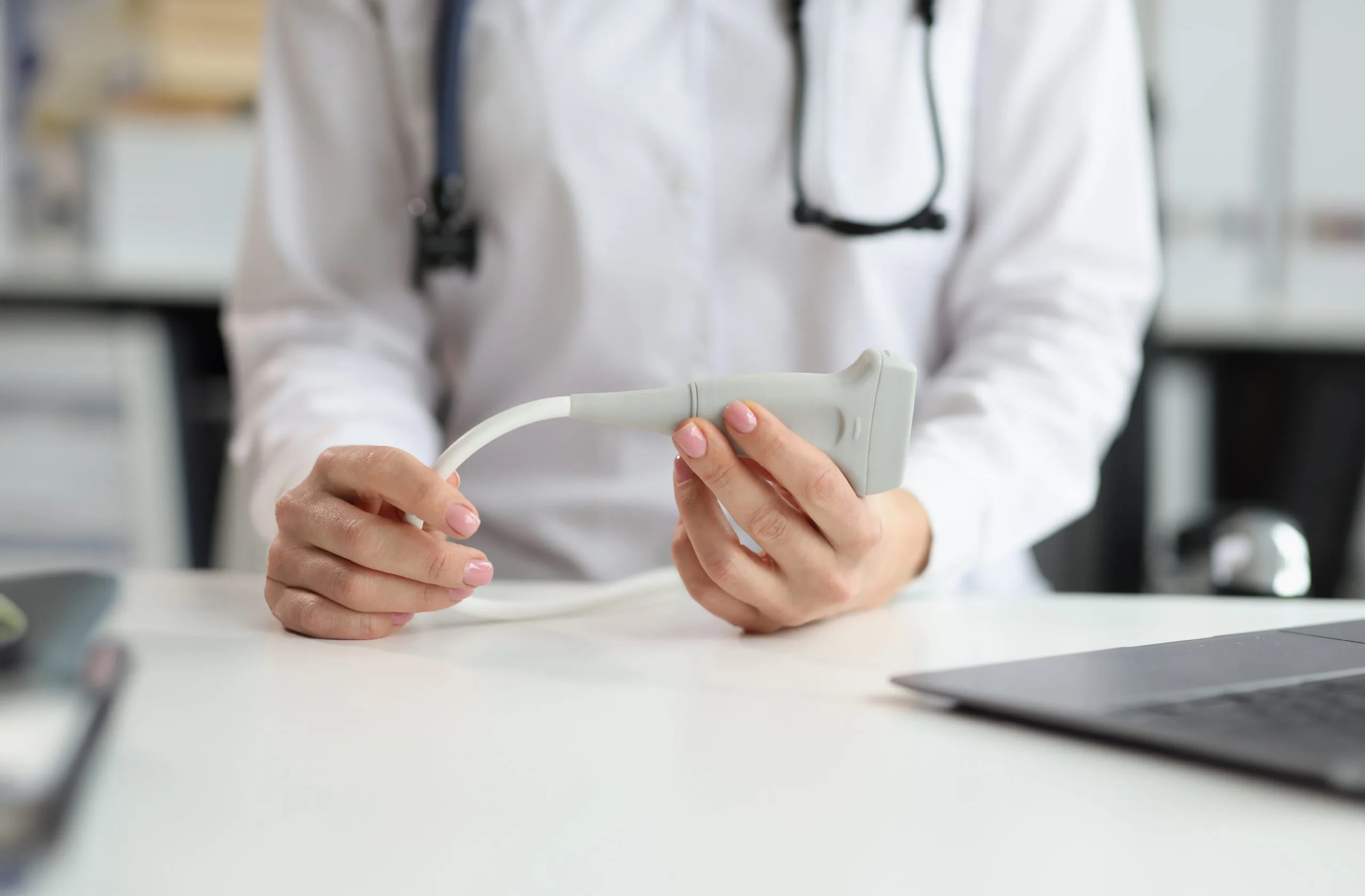
Ultrasound uses sound waves to create a live picture of the tissue under the skin. It does not use radiation. During the procedure, a small handheld probe rests on the skin while the surgeon works. The screen shows the tissue layers and the position of the instrument as it moves.
Depth control. The surgeon can confirm that fat is placed in the planned layer and kept away from deeper structures.
Live feedback. The image updates as the instrument moves, so adjustments happen in the moment.
Even distribution. Seeing where fat has already been placed helps spread small amounts evenly, which gives each graft a better blood supply.
Less guesswork. Visual confirmation supports the surgeon’s hands and judgment rather than replacing them.
Placing fat in thin, well-supported layers also supports graft survival. When more of the transferred fat survives, results tend to look smoother and last longer.
Ultrasound guidance is well established across medicine. Physicians use it to guide needles during nerve blocks, biopsies and vascular access. The principle is simple: when you can see the target, you can reach it more accurately. Plastic surgeons have applied the same idea to fat grafting, where precise depth matters.
For patients, ultrasound guidance adds very little to the experience. The probe is used while you are under anesthesia, so you will not notice it. It adds no incisions and no radiation. What it adds is a more controlled placement of your own fat.
Dr. Chris Devulapalli, a board certified, fellowship trained plastic surgeon, uses ultrasound guided fat transfer injections as part of his breast fat grafting approach at our Harrison office. If you are considering breast fat transfer, he can walk you through how image guidance fits your plan during a consultation.

About the Author
Dr. Devulapalli
October 19, 2026